Posted on

Vaccination and Titer Testing

Vaccination and Titer Testing.

Dogs and Cats

What we need to know

Professor Michael J. Day

BSc BVMS(Hons) PhD DSc DiplECVP FASM FRCPath FRCVS

School of Veterinary Sciences, University of Bristol

INTRODUCTION: Vaccination and Titer Testing

Dogs and Cats

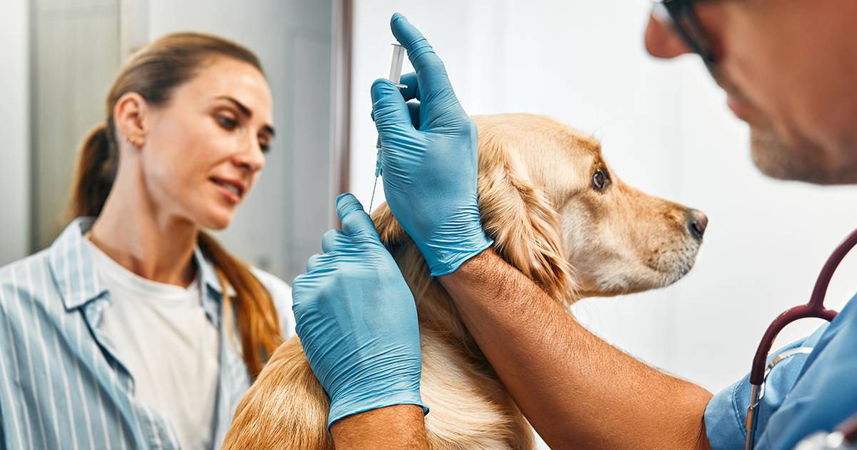
Vaccination delivery to dogs and cats has changed in the past decade. In response to questions over vaccine safety, guidelines groups have introduced new vaccination schedules that vaccine manufacturers have accommodated by introducing products with extended duration of immunity (DOI) and products with fewer antigenic components. The latest advance in vaccinology is the availability of simple in-practice test kits that demonstrate whether an individual animal has serological evidence of protection. These test kits can now inform decision-making about vaccination in practice. This presentation briefly reviews recommended vaccination schedules and focuses on the potential applications for in-house serological testing.

VACCINATION GUIDELINES: Vaccination and Titer Testing

Dogs and Cats

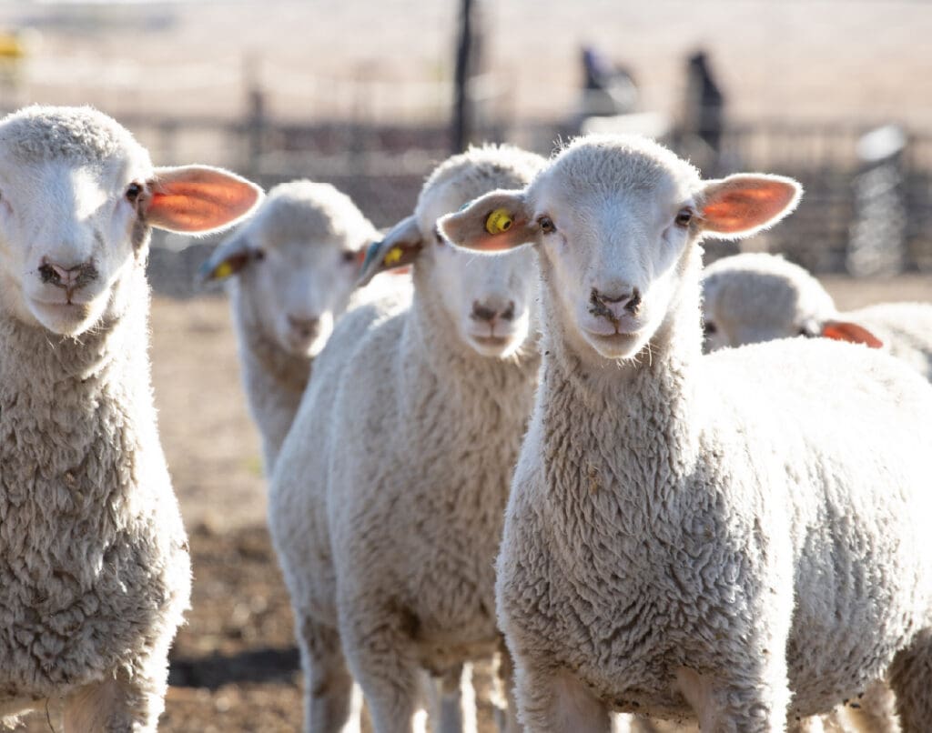
The WSAVA Vaccination Guidelines, as endorsed by BSAVA, consider vaccines as CORE or NON-CORE. CORE vaccines are those that every dog or cat should receive as they confer protection against diseases that are life-threatening or of significant morbidity. Even in developed countries, these diseases have not been eliminated, and the occurrence of regional outbreaks indicates the importance of maintaining herd immunity through vaccination. The CORE vaccine-preventable diseases of the dog are those induced by canine distemper virus (CDV), canine parvovirus-2 (CPV), and canine adenovirus-1 (CAV).

The CORE vaccine-preventable diseases of the cat are those induced by feline parvovirus (FPV), feline calicivirus (FCV), and feline herpesvirus-1 (FHV). WSAVA guidelines encourage the use of NON-CORE vaccines after making an appropriate benefit-risk analysis tailored to the individual pet’s lifestyle and risk of exposure. For that reason, in the UK, practitioners generally include vaccines to prevent leptospirosis as a CORE canine vaccine. They may include feline leukemia virus (FeLV) vaccines in the CORE feline vaccination schedule.

Modified live virus (MLV) or ‘infectious’ CORE vaccines should be administered to puppies and kittens from 8 – 9 weeks of age, with a second dose given 3 – 4 weeks later and a final dose between 14 – 16 weeks. This change ensures that all pups and kittens can mount at least a primary immune response to vaccination, even when maternally-derived antibody (MDA) persists to 12 weeks of age. Pups and kittens should also receive a booster CORE vaccine either 12 months after the final of the early life series or at 12 months of age.

Adult dogs and cats should receive MLV CORE vaccines no more frequently than every three years.

WSAVA recommendations also call for a triennial revaccination with FeLV. In the UK, most canine CORE vaccines on the market now carry a licensed 3-year DOI, and at least some FPV vaccines are licensed for triennial use. NON-CORE or ‘non-infectious’ vaccines other than FeLV must still be given annually to adult animals where these products are incorporated into an individualized vaccination program.

The overarching new concept in vaccination is that vaccines be delivered as one component of an ‘annual health check’ consultation that addresses all aspects of the health and well-being of the individual animal. Vaccination programs should be tailored to the requirements of the specific pet (individualized medicine) based on a thorough assessment of that animal’s lifestyle and risk factors. Most UK practitioners will currently administer triennial MLV CORE vaccines to the dog with an annual leptospirosis vaccine. Practitioners will also use a triennial program for feline CORE vaccines. Still, where an individual cat is perceived as having a higher risk for the feline infectious respiratory disease complex, that cat may receive annual FCV and FHV components, which are available as a separate combination product.

CORRELATES OF PROTECTION: Vaccination and Titer Testing

Dogs and Cats

For licensing studies, it is still necessary for animals to be used in experimental challenge studies in which vaccinated animals are challenged with the virulent form of a pathogen some time (e.g., 3 or 4 years) after vaccination to demonstrate either sterilizing immunity (failure of the pathogen to infect) or amelioration of clinical disease (infection occurs but the effects are limited). Studies over the decades have shown strong correlates of protection in such challenging studies.

For CDV, CPV, CAV, and FPV, the presence of serum antibodies that neutralize infectious viruses and prevent infection and disease provides a robust correlate of protection. This correlation is so strong that it is possible to state that the presence of serum antibodies to one of those viruses equates definitively with protective immunity. Some regulatory authorities are now beginning to accept seroprotection rather than an experimental challenge in modulating license claims. The presence of serum antibodies does not, however, provide a correlate of immunity for FCV and FHV protection. For respiratory pathogens such as FCV, mucosal secretory IgA provides a correlate of protection, but it is impossible to measure these antibodies routinely. For FHV, there is a stronger correlation between safety and cell-mediated immunity (CMI), but it is challenging to measure CMI on a routine basis.

GOLD STANDARD TESTS: Vaccination and Titer Testing

Dogs and Cats

Traditionally, two gold standard tests for CORE virus infections have measured the correlation between challenge immunity and seroprotection. These are the virus neutralization (VN) and haemagglutination inhibition (HAI) tests. A positive VN test indicates that serum from the animal contains antibodies that will neutralize infectious virus particles in vitro and prevent them from subsequently producing infection and cell damage (as assessed by the cytopathic effect on cells in vitro). A positive HAI test indicates that serum from the animal contains antibodies that will bind to infectious virus particles and neutralize the ability of that virus to cause agglutination of erythrocytes from particular animal species.

An excellent correlation exists between a positive VN test and protection against CDV, CPV, CAV, FPV, and rabies. There is also an excellent correlation between a positive HAI test and protection against CPV and FPV. For FCV, the positive VN test and protection correlation are considered only good to fair (as secretory IgA provides a better measure). For FHV, the positive VN test and protection correlation are only fair (as CMI is a better correlate of immunity).

Other serological test methods (e.g., ELISA or IFA-based) must, in turn, be correlated with the gold standard VN or HAI tests. Both of the in-house test systems that will be described below have had such correlation and validation with sera derived from animals in challenge studies tested by the
gold standards.

THE CONCEPT OF TITER: Vaccination and Titer Testing

Dogs and Cats

A titer measures the concentration of antibodies in a serum sample. Obtaining a titer involves performing an immunological test in which the serum sample is subjected to a series of doubling dilutions (that progressively reduce the antibody concentration). Each serum dilution is tested, and the highest dilution that gives an unequivocally positive reaction in the test provides the titer. The titer is the reciprocal of that dilution (e.g., a dilution of 1/10 provides a titer of 10).

Testing laboratories have traditionally provided a titer for serological tests of antibodies related to vaccine protection. The actual number may sometimes differ for the same sample tested by different laboratories. The number is relatively arbitrary as the titer is not a single defined
number but represents a range. For example, a sample with a titer of 10 indicates that the value is not less than 5 and not more than 20 (one doubling dilution above and below the titer). A sample with a titer of 1280 has a titer that is not less than 640 and not more than 2560. In
this regard, a titer of 640 from one laboratory is the same as one of 2560 from another testing laboratory.

For this reason, the WSAVA Vaccination Guidelines Group encourages practitioners to consider that any titer (above the cut-off for the gold standard test; typically 20 for HAI and 100 for VN) should be regarded as positive, and protection in challenge studies is still conferred throughout a broad spectrum of titers. For this reason, the new in-house test kits either provide a simple yes-no answer or a semiquantitative score, as antibodies above control levels correlate with protection (for those diseases defined above).

IN-HOUSE TEST KITS: Vaccination and Titer Testing

Dogs and Cats

Now, two companies produce in-house test kits for the determination of protective serum antibodies to CORE infectious diseases post-vaccination. Both test kit systems are simple to use, provide a rapid answer (protection or not) within 20 – 30 minutes, and are relatively inexpensive (costing around the same for testing as for revaccinating the animal). Several diagnostic laboratories have validated both test kit systems and correlated them with gold-standard tests. The test kits are the TiterCHEKTM system (manufactured by Synbiotics and now owned and distributed by Pfizer) and the VacciCheckTM system (produced by Biogal Laboratories).

The TiterCHEKTM system provides a yes-no (protected or not protected) answer for CDV and CPV. The VacciCheckTM system provides a semiquantitative score for serum antibody titers against CDV, CAV, and CPV. A feline VacciCheckTM system scores serum antibody titers against FPV, FCV, and FHV. The kits have excellent overall sensitivity (detecting samples with antibodies from those seropositive by gold standard) and specificity (detecting samples without antibodies from those seronegative by gold standard).

A set of excellent ‘YouTube’ videos produced by the US Charity ‘Maddie’s Fund’ is available on the web that provides very clear instructions on how to perform and interpret each of these test systems. Minor differences between the two systems are summarized below:

Vaccination and Titer Testing

APPLICATIONS OF IN-HOUSE TESTING

To Determine Puppy Protection and Detect Genetic Non-Responders: Using in-house test kits provides a simple measure of whether a puppy (CDV, CAV, CPV) or kitten (FPV) is protected after the initial series of early life vaccinations. This has the benefit of identifying animals that may not have responded to early life vaccination (particularly where a 14 – 16-week vaccine is not given) and may remain unprotected until the time of the 12-month booster. At this stage, a seropositive and protected animal may not require the 12-month booster and could go straight to a triennial CORE vaccination program.

WSAVA guidelines recommend the final CORE vaccination at 14 – 16 weeks.

The puppy can be tested 2 weeks after this vaccination (typically at 18 weeks). Seropositivity at this stage indicates that the pup has made an endogenous immune response to the vaccine, as there can be no MDA remaining at this time. A seronegative puppy at 18 weeks should be revaccinated (perhaps with an alternative product) and then tested 2 weeks later. A positive result indicates protection. A second negative result may indicate that the pup is either a ‘low responder’ or a ‘non-responder.’ Performing a gold standard test at this stage may show the low antibody titer typical of a low responder dog. Such an animal will be protected from clinical disease but not from infection.

Alternatively, the dog may lack antibodies and be a genetic non-responder incapable of ever making an immune response to that particular antigen.

Such dogs are, therefore, susceptible to infection and disease for life. Dogs of the Rottweiler breed have a higher proportion of genetic non-responders to parvovirus and rabies virus vaccines. Although non-responder rottweilers are no longer recognized in the US (the gene pool has been selected against them), they are still seen in Europe. Genetic non-responders generally cannot respond to one (rather than all) CORE vaccine antigens. The estimated prevalence of non-responders (US data) for CPV is 1 in every 1000 dogs and 1 in every 5000 dogs for CDV. CAV non-responders are rare (estimated < 1 in every 100,000 dogs).

A recent Danish study has evaluated seroconversion in a population of 135 pups aged between 8 weeks and 12 months. Most of these dogs will have finished an early life protocol (unlikely to have included a 14 – 16 week vaccine) but have not yet received a 12-month booster. The prevalence of
As determined by VacciCheckTM testing, 25.3% of this population were nonresponders for CPV, 20.7% for CAV, and 12.6% for CDV.

To Decide about the Vaccination of a ‘Lapsed’ Adult Dog

Much is currently made of revaccinating ‘lapsed’ adult or adult dogs adopted without a vaccination history. Most current data sheets for MLV CORE vaccines suggest that it is necessary to treat such animals as puppies and give two injections 3 – 4 weeks apart. Immunologically, an adult dog can be primed, immunized, and boosted from a single injection of MLV CORE vaccine, as there is no inhibitory MDA.

However, such dogs may not require vaccination at all, either because they have been previously vaccinated or, in some instances, have acquired natural immunity from field exposure to the virus. Owners may, therefore, be offered serology rather than automatic vaccination in this circumstance. An adult dog with serum antibodies to CDV, CAV, and CPV is protected already and does not require revaccination at that point. Similarly, a ‘lapsed’ or adopted adult cat with serum FPV antibody is protected and does not require that component of the vaccine at that time point.

To Minimize Risk in an Animal Previously Having an Adverse Reaction to a Vaccine

Adverse reactions of a broad spectrum are recognized post-vaccination in dogs and cats. The prevalence of these is low, and most have mild and transient effects. However, some (e.g., canine immune-mediated disease) are potentially life-threatening. If there is a suspicion that vaccination might have triggered a disease, then such animals should be subject to rigorous benefit-risk analysis before revaccination is considered. For CORE vaccine antigens, this decision is now made simpler by the availability of in-house serology. A dog with serum antibodies to CDV, CAV, and CPV does not require revaccination with MLV CORE vaccines, and serious consideration should be given to which NON-CORE products such an animal receives.

Serology Replacing Revaccination in an Annual Health Check

The Annual Health Check concept is gaining momentum in the US and Europe. So, too, is the adoption of triennial CORE revaccination schedules for adult animals. However, many US practices have continued in this rapidly changing arena. Instead of offering triennial CORE revaccination, these practices now provide the alternative of triennial serological testing using one of the in-house systems. Seropositive dogs (or cats seropositive for FPV) are not revaccinated with CORE vaccines as these are not required. NON-CORE vaccines may still be used annually, and FCV and FHV revaccination might be considered annually for cats at risk. Where this approach is used, the testing interval is reduced to annually for senior animals (dogs > 10 years and cats >15 years) to ensure that immunosenescence (aging of the immune system) is not an issue.

Management of Disease Outbreaks in Shelters

One of the most valuable applications of in-house serology has been managing infectious disease outbreaks in shelters, specifically for CDV, CPV, and FPV outbreaks. The ability to rapidly and cheaply test populations to identify animals that are protected or susceptible has allowed many animals to live that might otherwise have been euthanized as they were of unknown status.

In the face of a disease outbreak, all animals currently resident within the shelter should be tested. Those who are seropositive are protected and will not become infected or die. This protected population should be separated from low or negative responder animals, which should be isolated. The susceptible population should not be adopted out of the shelter until at least 2 weeks for CPV or FPV or until at least 6 weeks for CDV (reflecting the incubation periods of the diseases). The susceptible population might be retested after these intervals.

The second population to be considered is those animals that wish to enter the shelter. These should also be tested before considering admission. Seropositive animals may enter as they are protected from disease. Seronegative animals should be vaccinated and then ideally sent to foster homes. They should not be allowed to enter the shelter until they have seroconverted (when retested two weeks later).

This approach has proven successful in controlling infectious disease outbreaks in shelters. However, it is not applicable to outbreaks of the feline infectious respiratory disease complex, as serology is not correlated with protection.

FURTHER READING AND INFORMATION

http://www.wsava.org/VGG1.htm [WSAVA Vaccination Guidelines]

http://www.maddiesfund.org/ [videos on performing and interpreting in-house tests and use of
serology in control of outbreaks in shelters]

DiGangi BA, Gray LK, Levy JK, Dubovi EJ, Tucker SJ (2011) Detection of protective antibody titers
against feline panleukopenia virus, feline herpesvirus-1, and feline calicivirus in shelter cats using a
point-of-care ELISA. Journal of Feline Medicine and Surgery 13, 912-918.

Lund JD, Prior M, Madsen L (2012) Testing dogs for immunity against canine parvovirus, canine
distemper virus, and infectious canine hepatitis. Unpublished data.

Mazar SS, Dubovi EJ, Lavi Y, Lappin M (2009) Sensitivity, specificity, accuracy, and difference between positive and negative mean results of the ImmunoCombTM Feline VacciCheck antibody test kit for
feline calicivirus, rhinotracheitis, and panleukopenia. Unpublished data.

Mazar S, Larson L, Lavi Y (2009) Sensitivity-specificity-accuracy and difference between positive and negative mean results of the ImmunoCombTM Canine VacciCheck antibody test kit for canine
distemper, parvo and adenovirus. Unpublished data.

Waner T, Mazar S, Keren-Kornblatt (2006) Application of a dot enzyme-linked immunosorbent assay
for evaluation of the immune status to canine parvovirus and distemper virus in adult dogs before
revaccination. Journal of Veterinary Diagnostic Investigation 18, 267-270.

Posted on

Purdue Vaccination Studies

Purdue Vaccination Studies.

And Auto-antibodies

By Catherine O’Driscoll – Reprinted with permission from Dogs Naturally Magazine. Copyright (C) 2015 All Rights Reserved.

Purdue University School of Veterinary Medicine

A team at Purdue University School of Veterinary Medicine conducted several studies (1, 2) to determine if vaccines can cause changes in the immune system of dogs. These changes might lead to life-threatening immune-mediated diseases. The Purdue Vaccination Studies and Auto-antibodies. They conducted this research because concerns already existed. The Haywood Foundation sponsored it. They sought evidence that such changes in the human immune system might also be vaccine-induced. It found the evidence.

The vaccinated, but not the non-vaccinated, dogs in the Purdue studies developed autoantibodies to many of their biochemicals, including fibronectin, laminin, DNA, albumin, cytochrome C, cardiolipin, and collagen.

This means that the vaccinated dogs — ”but not the non-vaccinated dogs”– were attacking their fibronectin, which is involved in tissue repair, cell multiplication and growth, and differentiation between tissues and organs in a living organism.

Vaccines appear to be capable of removing cells’ natural intelligence.

The vaccinated Purdue dogs also developed autoantibodies to laminin, which is involved in many cellular activities, including cell adhesion, spreading, differentiation, proliferation, and movement. Vaccines, thus, appear to be capable of removing cells’ natural intelligence.

Autoantibodies to cardiolipin are frequently found in patients with the severe disease systemic lupus erythematosus, and also in individuals with other autoimmune diseases. Elevated anti-cardiolipin antibodies are significantly associated with clots within the heart or blood vessels, poor blood clotting, hemorrhage, bleeding into the skin, fetal loss, and neurological conditions.

The Purdue studies also found that vaccinated dogs developed autoantibodies against their collagen. About one-quarter of all the protein in the body is collagen. Collagen provides structure to our bodies, protecting and supporting the softer tissues and connecting them with the skeleton. No wonder Canine Health Concern’s 1997 study of 4,000 dogs showed many dogs developing mobility problems shortly after vaccination (noted in my 1997 book, What Vets Don’t Tell You About Vaccines).

DNM ACADEMY EXTRA: SAFER VACCINES & NOSODES

Dr. Dee Blanco will discuss the dangers of vaccinating your dog and why this alternative is so popular!

Not an Academy Member?

Get access to this course plus all the Academy has to offer for just $1

Perhaps most worryingly, the Purdue studies found that the vaccinated dogs had developed autoantibodies in their DNA. Did the alarm bells sound? Did the scientific community call a halt to the vaccination program? No. Instead, they stuck their fingers in the air, saying more research is needed to ascertain whether vaccines can cause genetic damage. Meanwhile, the study dogs were found in good homes, but no long-term follow-up has been conducted.

American Veterinary Medical Association (AVMA) Vaccine-Associated Feline Sarcoma Task Force

At around the same time, the American Veterinary Medical Association (AVMA) Vaccine-Associated Feline Sarcoma Task Force initiated several studies to find out why 160,000 cats in the USA develop terminal cancer each year at their vaccine injection sites. (3) The fact that cats can get vaccine-induced cancer has been acknowledged by veterinary bodies worldwide. Even the British Government acknowledged it through its Working Group. They were charged with looking into canine and feline vaccines (4), following pressure from Canine Health Concern. What do you imagine was the advice of the AVMA Task Force, veterinary bodies, and governments? “Carry on vaccinating until we find out why vaccines are killing cats and which cats are most likely to die.”

To mitigate the problem in America. They’re vaccinating cats in the tail or leg so they can amputate when cancer appears. Great advice if it’s not your cat amongst the hundreds of thousands on the “oops” list.

In August 2003, the Journal of Veterinary Medicine published an Italian study. The study shows that dogs also develop vaccine-induced cancers at their injection sites.

But other species are okay. Wrong. In August 2003, the Journal of Veterinary Medicine published an Italian study. This study shows that dogs also develop vaccine-induced cancers at their injection sites. (5) We already know that vaccine-site cancer is a possible sequel to human vaccines, too. Since the Salk polio vaccine was said to carry a monkey retrovirus (from cultivating the vaccine on monkey organs) that produces inheritable cancer. The monkey retrovirus SV40 keeps turning up in human cancer sites.

No one warns pet owners before their animals are subjected to an unnecessary booster.

It is also widely acknowledged that vaccines can cause a fast-acting, usually fatal, disease called autoimmune hemolytic anemia (AIHA). Without treatment, and frequently with treatment, individuals can die in agony within a matter of days. Merck, a multinational vaccine manufacturer, states in The Merck Manual of Diagnosis and Therapy that autoimmune hemolytic anemia may be caused by modified live-virus vaccines, as do Tizard’s Veterinary Immunology (4th edition) and the Journal of Veterinary Internal Medicine. (6) The British Government’s Working Group, despite being staffed by vaccine-industry consultants who say they are independent, also acknowledged this fact. However, no one warns pet owners before their animals are subjected to an unnecessary booster, and very few owners are told why after their pets die of AIHA.

A Wide Range of Vaccine-induced Diseases: The Purdue Vaccination Studies and Auto-antibodies

Our 1997 survey also found some worrying correlations between vaccine events and the onset of arthritis. Research in the human field compounded our concerns.

For example, the New England Journal of Medicine reported that isolating the rubella virus from affected joints in children vaccinated against rubella is possible. It also told of the isolation of viruses from the peripheral blood of women with prolonged arthritis following vaccination. (7)

Then, in 2000, CHC’s findings were confirmed by research. The research showed that polyarthritis and other diseases like amyloidosis, which affects organs in dogs, were linked to the combined vaccine given to dogs. (8) There is a vast body of research, despite the lack of funding from the vaccine industry, to confirm that vaccines can cause a wide range of brain and central nervous system damage. Merck states in its Manual that vaccines (i.e., its products) can cause encephalitis and brain inflammation/damage. In some cases, encephalitis involves lesions in the brain and throughout the central nervous system. Merck states, “Examples are the encephalitides following measles, chickenpox, rubella, smallpox vaccination, vaccinia, and many other less well-defined viral infections.”

When the dog owners who took part in the CHC survey reported that their dogs developed short attention spans, 73.1% did so within three months of a vaccine event. The same percentage of dogs were diagnosed with epilepsy within three months of a shot (but usually within days). We also found that 72.5% of dogs whose owners considered nervous and of a worrying disposition first exhibited these traits within the three-month post-vaccination period.

Encephalitis, incidentally, is a disease that can manifest across the scale from mild to severe and can also cause sudden death.

I want to add, for the sake of Oliver, my friend who suffered from paralyzed rear legs and died shortly after a vaccine shot, that “paresis” is listed in Merck’s Manual as a symptom of encephalitis. This is defined as muscular weakness of a neural (brain) origin, which involves partial or incomplete paralysis resulting from lesions at any level of the descending pathway from the brain. Hind limb paralysis is one of the potential consequences. Encephalitis, incidentally, is a disease that can manifest across the scale from mild to severe and can also cause sudden death.

Organ failure must also be suspected shortly after a vaccine event.

Dr Larry Glickman, who spearheaded the Purdue research into post-vaccination biochemical changes in dogs, wrote in a letter to Cavalier Spaniel breeder Bet Hargreaves:

“Our ongoing studies of dogs show that following routine vaccination, there is a significant rise in the antibodies dogs produce against their tissues. Some of these antibodies have been shown to target the thyroid gland and connective tissue, such as those found in the valves of the heart, red blood cells, DNA, etc. I believe that the heart conditions in Cavalier King Charles Spaniels could result from repeated immunizations by vaccines containing tissue culture contaminants that cause a progressive immune response directed at connective tissue in the heart valves. The clinical manifestations would be more pronounced in dogs with a genetic predisposition, although the findings should generally apply to all dogs regardless of their breed.”

I must mention here that Dr Glickman believes that vaccines are a necessary evil, but safer vaccines must be developed.

Vaccines Stimulate an Inflammatory Response:

The word “allergy” is synonymous with “sensitivity” and “inflammation.” It should, by rights, also be synonymous with the word “vaccination.” Vaccines do this by sensitizing (rendering an individual allergic) and forcing them to develop antibodies to fight a disease threat. In other words, as is acknowledged and accepted, the body will respond with inflammation as part of the vaccine process. This may be temporary or longstanding.

Holistic doctors and veterinarians have known this for at least 100 years. They discuss various inflammatory or “-itis” diseases that arise shortly after a vaccine event. Vaccines plunge many individuals into an allergic state. Again, this is a disorder that ranges from mild to suddenly fatal. Anaphylactic shock is the culmination: it’s where an individual has a massive allergic reaction to a vaccine and will die within minutes if adrenaline or its equivalent is not administered.

Some individuals are genetically not well placed to withstand the vaccine challenge.

These are the people (and animals are “people,” too) who have inherited faulty B and T cell functions. B and T cells are components within the immune system that identify foreign invaders, destroy them, and hold the invaders in memory so that they cannot cause future harm. However, where inflammatory responses are concerned, the immune system overreacts and causes unwanted effects such as allergies and other inflammatory conditions.

Merck warns in its Manual that patients with, or from families with, B and/or T cell immunodeficiencies should not receive live-virus vaccines. Stating that it is due to the risk of severe or fatal infection. Elsewhere, it lists features of B and T cell immunodeficiencies such as food allergies, inhalant allergies, eczema, dermatitis, neurological deterioration, and heart disease. To translate, people with these conditions can die if they receive live-virus vaccines. Their immune systems are not competent enough to guarantee a healthy reaction to the viral assault from modified live-virus vaccines.

Modified live-virus (MLV) vaccines replicate in the patient until an immune response is provoked. If a defense isn’t stimulated, the vaccine replicates until it gives the patient the disease it intended to prevent.

Alternatively, a deranged immune response will lead to inflammatory conditions such as arthritis, pancreatitis, colitis, encephalitis, and any number of autoimmune diseases, such as cancer and leukemia, where the body attacks its cells.

A new theory, stumbled upon by Open University student Gary Smith, explains what holistic practitioners have been saying for a long time. Here is what a few of the holistic vets have said about their patients:

Dr Jean Dodds:

“Many veterinarians trace the present problems with allergic and immunologic diseases to the introduction of MLV vaccines…” (9)

Christina Chambreau, DVM:

“Routine vaccinations are probably the worst thing that we do for our animals. They cause all types of illnesses, but not directly to where we would relate them definitely to be caused by the vaccine.” (10)

Martin Goldstein, DVM:

“I think that vaccines…are leading killers of dogs and cats in America today.”

Dr Charles E. Loops, DVM:

“Homoeopathic veterinarians and other holistic practitioners have maintained for some time that vaccinations do more harm than they provide benefits.” (12)

Mike Kohn, DVM:

“In response to this [vaccine] violation, there have been increased autoimmune diseases (allergies being one component), epilepsy, neoplasia [tumors], as well as behavioral problems in small animals.” (13)

A Theory on Inflammation:

Gary Smith explains what observant healthcare practitioners have been saying for a long time, but perhaps they’ve not understood why their observations led them to say it. His theory, incidentally, is causing a massive stir within the inner scientific sanctum. Some believe that his theory could lead to a cure for many diseases, including cancer. For me, it explains why the vaccine process is inherently questionable.

Gary was studying inflammation when he struck upon a theory so extraordinary that it could have implications for the treatment of almost every inflammatory disease, including Alzheimer’s, Parkinson’s, rheumatoid arthritis, and even HIV and AIDS.

Gary’s theory questions the received wisdom that when a person gets ill, the inflammation around the infected area helps it heal. He claims that, in reality, inflammation prevents the body from recognizing a foreign substance and, therefore, serves as a hiding place for invaders. The inflammation occurs when at-risk cells produce all receptors (angiotensin II type I receptors). He says At1 has a balancing receptor, At2, which is supposed to switch off inflammation, but this does not happen in most diseases.

“Cancer has been described as the wound that never heals,” he says. “All successful cancers are surrounded by inflammation. This is commonly thought to be the body’s reaction to fight the cancer, but this is not the case.

“The inflammation is not the body trying to fight the infection. It is the virus or bacteria deliberately causing inflammation to hide from the immune system [author’s emphasis].” (14)

If Gary is correct, the inflammatory process commonly stimulated by vaccines is not, as hitherto assumed, a necessarily acceptable sign. Instead, it could be a sign that the viral or bacterial component, or the adjuvant (which contains foreign protein and is seen as an invader by the immune system), in the vaccine, is winning by stealth.

If Gary is correct in believing that the inflammatory response is not protective but a sign that invasion is taking place under the cover of darkness, vaccines are certainly not the friends we thought they were. They are undercover assassins working on behalf of the enemy, and vets and medical doctors are unwittingly acting as collaborators. Worse, we animal guardians and parents are paying doctors and vets to betray our loved ones, unwittingly.

Potentially, vaccines are the stealth bomb of the medical world. They catapult invaders inside the castle walls, where they can wreak havoc, with none of us any the wiser. So rather than experiencing frank viral diseases such as the flu, measles, mumps, and rubella (and, in the case of dogs, parvovirus and distemper), we are allowing the viruses to win anyway—but with cancer, leukemia, and other inflammatory or autoimmune (self-attacking) diseases taking their place.

The Final Insult: The Purdue Vaccination Studies and Auto-antibodies

All 27 veterinary schools in North America have changed their protocols for vaccinating dogs and cats (15). However, vets in practice are reluctant to follow these changed protocols, and official veterinary bodies in the UK and other countries are ignoring the following facts.

Dogs’ and cats’ immune systems mature fully at six months. If a modified live-virus vaccine is given after six months of age, it produces immunity. This is suitable for the pet’s life. If another MLV vaccine is given a year later, the antibodies from the first vaccine neutralize the antigens of the second vaccine. There is little or no effect. The liter is not “boosted,” nor are more memory cells induced.

Annual boosters are unnecessary, subjecting the pet to potential risks such as allergic reactions and immune-mediated hemolytic anemia.

In plain language, veterinary schools in America, plus the American Veterinary Medical Association, have looked at studies to show how long vaccines last. They have concluded and announced that annual vaccination is unnecessary. (16-19)

Further, they have acknowledged that vaccines are not without harm. Dr Ron Schultz, head of pathobiology at the University of Wisconsin and a leading light in this field, has said this politely to his veterinary colleagues since the 1980s. I’ve been saying it for the past 12 years. But change is so long in coming, and, in the meantime, hundreds of thousands of animals are dying unnecessarily every year.

The good news is that thousands of animal lovers (but not enough) have heard what we’ve been saying. Canine Health Concern members worldwide use real food as Nature’s supreme disease preventative. Eschewing processed pet food and minimizing the vaccine risk. Some of us, including me, have chosen not to vaccinate our pets at all. Our reward is healthy and long-lived dogs.

It has taken but one paragraph to tell you the good and simple news. The gratitude I feel daily when I embrace my healthy dogs stretches from the center of the Earth to the Universe and beyond.

Endnotes: The Purdue Vaccination Studies and Auto-antibodies

  • 1. “Effects of Vaccination on the Endocrine and Immune Systems of Dogs, Phase II,” Purdue University, November 1, 1999, at http://www.homestead.com/vonhapsburg/haywardstudyonvaccines.html.
  • 2. See www.vet.purdue.edu/epi/gdhstudy.htm.
  • 3. See http://www.avma.org/vafstf/default.asp.
  • 4. Veterinary Products Committee (VPC) Working Group on Feline and Canine Vaccination, DEFRA, May 2001.
  • 5. JVM Series A 50(6):286-291, August 2003.
  • 6. Duval, D. and Giger, U. (1996). “Vaccine-Associated Immune-Mediated Hemolytic Anemia in the Dog,” Journal of Veterinary Internal Medicine 10:290-295.
  • 7. New England Journal of Medicine, vol.313,1985.
  • See also Clin Exp Rheumatol 20(6):767-71, Nov-Dec 2002.
  • 8. Am Coll Vet Intern Med 14:381,2000.
  • 9. Dodds, Jean W., DVM, “Immune System and Disease Resistance,” at http://www.critterchat.net/immune.htm.
  • 10. Wolf Clan magazine, April/May 1995.
  • 11. Goldstein, Martin, The Nature of Animal Healing, Borzoi/Alfred A. Knopf, Inc., 1999.
  • 12. Wolf Clan magazine, op. cit.
  • 13. ibid.
  • 14. Journal of Inflammation 1:3,2004, at http://www.journal-inflammation.com content/1/1/3.
  • 15. Klingborg, D.J., Hustead, D.R. and Curry-Galvin, E. et al., “AVMA Council on Biologic and Therapeutic Agents’ report on cat and dog vaccines”, Journal of the American Veterinary Medical Association 221(10):1401-1407, November 15,2002, http://www.avma.org/policies/vaccination.htm.
  • 16. ibid.
  • 17. Schultz, R.D., “Current and future canine and feline vaccination programs,” Vet Med 93:233-254,1998.
  • 18. Schultz, R.D., Ford, R.B., Olsen, J. and Scott, P., “Titer testing and vaccination: a new look at traditional practices,” Vet Med 97:1-13, 2002 (insert).
  • 19. Twark, L. and Dodds, W.J., “Clinical application of serum parvovirus and distemper virus antibody titers for determining revaccination strategies in healthy dogs,” J Am Vet Med Assoc 217:1021-1024,2000.
Posted on

Mechanism of Transfer Factor

Mechanism of Transfer Factor.

Balancing T-Helper Cell Activity

Mechanism of Transfer Factor

Question: How can Transfer Factor change a predominantly TH-2 Immune System to a TH-1 predominant System so rapidly?

Answer: In the case of Transfer Factor, clinicians have been well ahead of scientists and immunologists. Dr. Sherwood Lawrence, lead immunologist at New York University, discovered Transfer Factor in 1949. Though at the time, his discovery went no further than the library shelf. Now, there are an estimated 3,000 references to Transfer Factor in the literature worldwide. There are some on the adjuvants as well.

It has been a puzzle as to how transfer factors could have such rapid action if they depended upon the new production of T-helper cells from an exhausted immune system. Here is the mechanism: Eomesodermin.

T-Helper Cells develop along two different lines – TH-1 and TH-2.

TH-1 predominance modulates cell-mediated immunity and produces cytokines: IL-2, IFN-gamma, and TNF-alpha. TH-2 cells modulate humoral immunity, or antibody production, and produce IL-4, IL-5, IL-6, IL-10, and IL-13. TH-1 predominance increases the body’s ability to defend itself against viruses, fungi, parasites, cancer, and intracellular organisms.

TH-2 dominant states, with decreased cellular immunity and increased humoral immunity, tend to manifest as Allergies, Chronic Sinusitis, Atopic eczema, and Asthma. And in systemic autoimmune conditions such as CFS (now observed in horses), Lupus (SLE), Mercury-induced autoimmunity, Chronic Giardiasis, Chronic Candidiasis, viral infections, Ulcerative Colitis, and Cancer.

Transfer Factor augments cell-mediated immunity and modifies a TH-2 dominant state to TH-1 dominance.

If the TH-1 response has resulted in an inadequate attempt by the immune system to fight off and eliminate an infection or neoplasia, then Transfer Factor would augment that process and be effective in many cases. Clinically, one sees this in Crohn’s Disease, Multiple Sclerosis, and Chronic Lyme Disease. In these cases, Transfer Factors help and accentuate the efforts of an already TH-1 dominant immune system.

Mechanism of Transfer Factor: From SCIENCE, p. 1041, Vol. 302, Nov. 2003

“The attributes of “Helper” T-cells are governed by two master transcriptional regulators, GATA-3 and T-bet, which coordinate the expression of specific cytokine genes. However, neither appears critical to the CD8+ T-cell function, leading Pearce et al to explore other pathways that might specify the transcriptional program in these cells. The trail ended in the identification of Eomesodermin (Eomes), a T-box transcription factor related to T-bet and already characterized as a key regulator of mesodermal differentiation. Eomes was specifically up-regulated in activated CD8+ cells [one of TF’s actions], and forced cellular expression of the factor conferred CD8+ characteristics on cells that had already differentiated into TH-2 [T-helper type two] cells. [It was found that] Disruption of expression significantly reduced the CD8 cytolytic program and interferon Gamma expression.

Thus, Eomes cooperates with T-bet in regulating distinct aspects of cell-mediated immunity.”

Mechanism of Transfer Factor: Additional note

This is the mechanism of the transfer factor that increases the effectiveness of a TH-1 predominant immune system and influences change from a TH-2 state to a TH-1 state. In the past, we have not had an explanation of HOW TF affected the immune capability so rapidly. It is one thing to realize that new production T-helper cells could be configured into the TH-1’s, but with a marked effect in 48 hours and a height of effect in five days by TF, it seemed too fast – it takes ten days to two weeks for the production, maturation and configuration of T-cells. Now we see that the Eomes prompted by Transfer Factor while in circulation can convert TH-2s to TH-1s.

This is the point – Eomes was specifically up-regulated in activated CD8+ cells [one of TF’s actions], and forced cellular expression of the factor conferred CD8+ characteristics on cells that had already differentiated into TH-2.

Posted on

Immune Modulating Supplements

Immune Modulating Supplements.

Ramaekers Nutrition Immune System Enhancement

Priming the immune system to respond appropriately to natural infection and antigens from vaccines is what the Ramaekers Nutrition Immune Modulating supplements are indicated for. Our studies in several species, found under News & Articles, reveal how vaccines given along with our supplements induce a quicker, higher, and more long-lasting antibody titer.

Immune modulation is a therapy concept that encourages a more balanced immune response in the body. It up-regulates the immune cells as needed and, just as important, down-regulates the immune response when needed. The Innate Intelligence of the body strives for this balance at all times. Our unique combination of ingredients allows this to happen without side effects.

Immune Modulating Supplements

One of the main indications of our immune supplements is that they allow for fewer vaccines overall. Yet provide the opportunity for more long-lasting protection. Immunotherapy can also be used to enhance the immune response to vaccines and natural infections. This approach particularly targets the 10% to 20% of the population identified as poor responders. That is to say, those individuals who fail to produce adequate levels of protective antibodies. Our studies found in News & Articles demonstrate that both of these actions do happen.

Also interesting is that when taking these types of immune active ingredients, we don’t observe clinically any increased allergies or adverse reactions in either healthy or sick patients. These products are so safe that they are given to newborns of all domesticated species.

So the message is, to improve the response to necessary vaccines, establish higher levels and more long-lasting levels of protection, need vaccine updates less often, and improve protection against and response to natural infection, it is advised to use Ramaekers Immune Formulas, available in non-ruminating and ruminating forms, see bovine products, and other livestock products.

Posted on

Next Step In Immunotherapy

Next Step in Immunotherapy.

Transfer Factor

Next Step In Immunotherapy: Updated: Feb 19, 2024
Original: Feb 01, 2003

By Kenneth Marcella, DVM

DVM360 MAGAZINE

Proposed advances

Immunotherapy was predicted to be one of the most rapidly expanding areas of medical science in this decade.

Proposed advances in our ability to manipulate the protection offered by the body’s own immune system would make humans and animals far healthier and promise to increase both the length and quality of life.

Immune agents would be our new defenses against microorganisms that no longer respond to antibiotics, and this new field would subdue diseases from allergies to cancer.

Transfer Factor: The long-awaited next step in immunotherapy

To date, however, those advances have been slow to come. We have been unable to manipulate the body’s defenses as planned, and we have few effective immune stimulants.

Positive research data has been piling up concerning the most promising immune agent in years, though immunologists may be finally making good on their earlier predictions.

As this new immune compound is being called transfer factor, it may be the long-awaited “next step,” and it may be everything promised.

Richard Bennett, Ph. D., an infectious disease immunologist, writes, “Our ability to create a really healthy immune system represents the greatest potential gains in health in the world.”

After all, the immune system allows humans and animals to recognize and remember potentially harmful foreign substances such as bacteria and viruses. It will then enable us to respond to these threatening invaders in our systems.

Devastatingly familiar

The consequences of suppressed or damaged immune system conditions are devastatingly familiar to veterinarians.

Arab foals with combined immunodeficiency complex and older horses with chronic laminitis due to Cushing’s disease-related effects are but two such examples. Respiratory problems in young foals, allergies, skin infections, and hoof wall diseases are other problems that can also be related to immune system dysfunction.

A new agent that would vastly improve horses’ immune function would warrant some attention. Transfer factor promises to be this new agent. It is a colostrum component produced to be used as a powder added to the diet.

Structure and function

To understand what this colostral derivative may potentially mean to veterinarians and horses, it is necessary to briefly review the structure and function of the immune system.

The body has two principal immune defense systems: humoral and cellular. B-lymphocytes in the liver, spleen, lymph nodes, and bone marrow produce plasma cells that, in turn, produce antibodies in the gamma globulin fraction. These immunoglobulins can potentially recognize vast numbers of antigens. This is the basis of humoral immunity.

Humoral immunoglobulins are primarily designed to fight bacterial infections. Lymphocytes that populate the thymus become responsible for cellular immunity. These cells produce structures called lymphokines that mediate delayed hypersensitivity or allergic reactions. They are accountable for rejecting transferred foreign tissue and recognizing and rejecting tumor cells.

The cellular immunity system defends against infections caused by viruses, fungi, and some types of bacteria and cancers. Transfer factor stimulates both portions of the immune system.

In the beginning

In 1949, Dr. H. Sherwood Lawrence, a researcher working on tuberculosis, found that he could transfer immunity between patients using fractionated white blood cells. The key ingredient was a part of the lymphocyte cell, and Lawrence called this component “transfer factor.”

This discovery was not actively pursued for nearly 30 years until the late ’80s. At that time, colostrum and milk were discovered to contain significant amounts of transfer factor. The exact mechanism of action of the transfer factor has never been determined, but it is now known that the transfer factor is a lymphokine. The two most notable lymphokines are interferon and interleukin.

These lymphokines are protein messengers thought to be released by antigen-sensitized lymphocytes. They play a role in macrophage activation, lymphocyte transformation (precursor cells becoming B and T cells), and cell-mediated immunity. Transfer factor is one of the most potent messengers and has three distinct effects on the immune system.

Recognizes antigens

Transfer Factor: The long-awaited next step in immunotherapy helps the body recognize antigens.

Dr. M. Metz, a veterinarian,  points out that 200 mg (one capsule) of transfer factor has the potential to recognize at least 100,000 pathogens. Metz adds that not only can the transfer factor be specific to an individual antigen that a lymphocyte is exposed to, but the “transfer factor can also stimulate a multivalent response.”

In this type of response, the transfer factor activates lymphocytes to several strains of an organism.

“This is the really exciting part of the transfer factor from a practicing veterinary standpoint,” says Metz.

Research has found that exposing cattle to various bacteria and viruses can produce a transfer factor that stimulates immunity to related strains of bacteria and viruses that are much more pathogenic to other species.

“The other really exciting aspect of transfer factor,” says Metz, “is the time sequence.”

Most types of delayed hypersensitivity immunity, such as that seen with vaccine use, take 10 to 14 days to develop. Metz says the transfer factor activates that same immunity in 24 hours!

Natural killer cells

Transfer factor is also a natural killer cell inductor. These nonspecific attack cells seek out and destroy infected or malignant cells and cells infected by viruses.

Transfer factor increases natural killer cell activity five times over typical rates, and it is non-species specific. This aspect of the transfer factor is believed to be related to the significant improvements seen in certain cancer patients who have taken this product. Multiple sclerosis patients have also shown improvements.

The transfer factor in cats, dogs, horses, cows, and humans is structurally and functionally identical. This has helped in producing this product since cows can produce large quantities of colostrum that is then used to extract transfer factors.

Suppresses (down-regulation) immune function

Transfer Factor: The long-awaited next step in immunotherapy also suppresses immune function.

Paradoxically, the same product can stimulate and suppress immune function, but transfer factor function depends on the specific antigens and the immune response status. Transfer factor can stimulate the release of T suppressor cells when “down” regulation is necessary due to overactivity. Autoimmune diseases, chronic obstructive pulmonary disease (COPD), and allergic reactions are situations where the body’s own immune response has overresponded to antigenic stimulation. The transfer factor works in these situations because it can slow down this overactive response.

The next step in immunotherapy: Transfer Factor enhances the body’s ability to combat bacteria and viruses.

Obvious advantages

While discussions of the immune system tend to be pretty technical, the practical advantages of a potent new immune-stimulating treatment are apparent.

The ability to stimulate the horse’s body to attack and destroy bacteria and viruses will reduce the amount and types of antibiotics that veterinarians may need to use. It is important to try to retain those antibiotics available to veterinarians and to use them to maintain their effectiveness for as long as possible.

If veterinarians can stimulate a better immune response to respiratory bacteria, skin pathogens, and various viruses, then the need to use antibiotics is lessened. If the transfer factor can produce such boosts in immunity in 24 hours, then the potential for use as a pre-travel protectant or a post-exposure treatment is tremendous.

Horses suffering from other diseases, such as Cushing’s disease, laminitis, colitis, cancers ranging from sarcoids to melanomas, and reproductive conditions such as chronic metritis, may all benefit from transfer factor use. This product may indeed be the long-awaited next step, and the field of immunotherapy may finally fulfill its promise.

Posted on

Enhancing Embryo Quality

Enhancing Embryo Quality.

Embryo Quality Characteristics with Superovulated Cows Treatment with Nutrition

Horizons Nutrition Factor Immune Product (NHNFI)
G.H.L. Marquezinia, V.R.G. Mercadantea, M.M. Wardb, A.R. Spellc, J. Carterc, N. Paton, G. C. Lamba

  • North Florida Research and Education Center, University of Florida, Marianna, FL;
  • Nutrition Horizons, Lewisburg, OH;
  • Applied Reproduction Resources, Daphne, AL